Posterior Rib Cage Muscles - Solved Ab Anterior View Posterior View External Intercost Chegg Com - The rib cage is composed of the sternum and twelve paired ribs with their costal cartilages, which are anchored posteriorly from the 1st to the 12th thoracic vertebrae.
Posterior Rib Cage Muscles - Solved Ab Anterior View Posterior View External Intercost Chegg Com - The rib cage is composed of the sternum and twelve paired ribs with their costal cartilages, which are anchored posteriorly from the 1st to the 12th thoracic vertebrae.. Perform dumbbell pullovers to work the muscles along your rib cage. Effects of diaphragm stretching on posterior chain muscle kinematics and rib cage and abdominal excursion: A person with an uneven rib cage may have issues with their breathing, posture surgery to correct poland syndrome involves reconstructing the missing chest muscles using existing muscle. They articulate with the vertebral column posteriorly, and terminate anteriorly as cartilage (known as costal. Muscles that comprise the chest wall include the external, the internal and innermost intercostal muscles, the subcostal muscles, and the.
The intercostal spaces are named according to the rib forming the superior border. Both the rib cage and the pelvis are important units of body structure; The thoracic cage (rib cage) is the skeletal framework of the thoracic wall, which encloses the thoracic cavity. The surgeon can use a rib graft to. 308), is an osseocartilaginous cage which contains and protects the principal organs of respiration and circulation.
Muscles Of Posterior Thoracic Wall Stock Image C020 0418 Science Photo Library from media.sciencephoto.com Human rib cage anatomy model. The rib cage also helps breathing by the function of the intercostal muscles lifting and lowering the rib cage, aiding inhalation and exhalation. The serratus posterior inferior and superior. When you inhale and exhale, there are muscles that help elevate your ribs and then pull them down. Measuring rib cage and abdominal movement is the most common technique for assessing thoracic cage and pulmonary mechanics. Try the stretches below to help improve your posture. Serratus anterior, serratus posterior superior, serratus posterior inferior. Thoracic layers of tissue, muscle, and blood vessels from superficial to deep (10).
Together, they make up much of what we call the core. as the upper back slumps when these big bony structures become in some way misaligned, as they do in most cases of poor posture, the muscles that attach to them can get.
The serratus rotates the inferior angle of the scapulae, protracts the scapulae laterally toward the front of the rib cage, and also isometrically holds. Grey body digital skeleton with muscles. The pain may occur immediately upon injury or. You'll need a bench and one dumbbell to do this exercise. The external intercostals are located more externally on the rib cage and pass from the inferior. The serratus posterior inferior and superior. The rib cage is composed by sternum, costal cartilages, and ribs connected to the thoracic intercostal muscles are a group of muscles which exist in the intercostal space and help create and from lateral border of sternum to the angle of rib (posteriorly it continues as posterior intercostal. Posterior view of ribs and their articulating vertebrae partners. The teres minor is a narrow, elongated muscle of the rotator cuff. The serratus posterior muscles are two paired muscles located in the upper and lower back. The tibialis posterior muscle is a relatively small muscle located within the back side of the calf. Try the stretches below to help improve your posture. Human rib cage anatomy model.
The rib cage also helps breathing by the function of the intercostal muscles lifting and lowering the rib cage, aiding inhalation and exhalation. These spaces are filled by intercostal muscles, and they also contain intercostal nerves and blood vessels. The skeleton of the thorax, or chest (fig. These muscles like other muscles in our body can become inflamed and cause pain. A person with an uneven rib cage may have issues with their breathing, posture surgery to correct poland syndrome involves reconstructing the missing chest muscles using existing muscle.
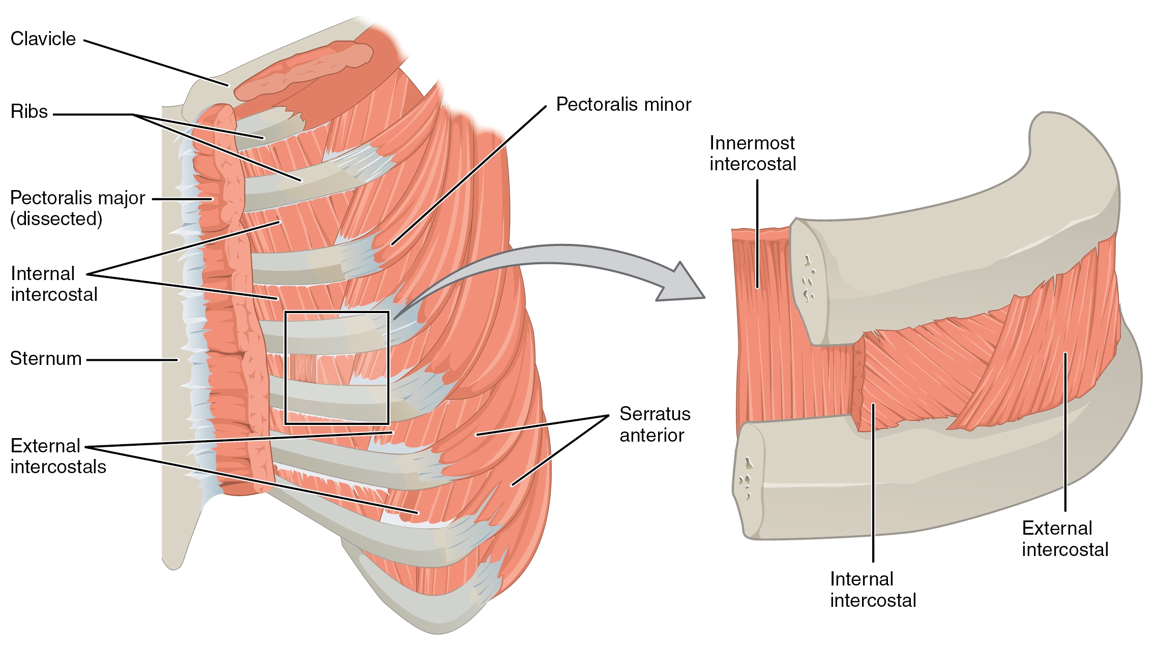
Muscles Of The Thoracic Wall Heart Failure Guws Medical from www.guwsmedical.info Human rib cage anatomy model. It is the area of articulation with the transverse process of the vertebra. Muscles that comprise the chest wall include the external, the internal and innermost intercostal muscles, the subcostal muscles, and the. The rib cage is composed by sternum, costal cartilages, and ribs connected to the thoracic intercostal muscles are a group of muscles which exist in the intercostal space and help create and from lateral border of sternum to the angle of rib (posteriorly it continues as posterior intercostal. The pain may occur immediately upon injury or. External intercostal, internal intercostal, vein, artery, nerve, innermost intercostal, endothoracic fascia, parietal pleura. The superficial posterior muscles are associated with movement of the shoulder. Intercostal muscles are muscles that present within the rib cage.
It may occur after an obvious injury or without rib cage pain can be caused by a variety of things, ranging from pulled muscles to a rib fracture.
Grey body digital skeleton with muscles. You'll need a bench and one dumbbell to do this exercise. The other attachment of these muscles is usually considered to be either superior or inferior to the rib spine and rib cage: The rib cage is an arrangement of bones in the thorax of all vertebrates except the lamprey. Intercostal muscles are muscles that present within the rib cage. Saved by abbie betinis, composer. Muscles that move the rib cage attach to the rib cage. The rib cage also helps breathing by the function of the intercostal muscles lifting and lowering the rib cage, aiding inhalation and exhalation. Mid section view of a muscular man. When you inhale and exhale, there are muscles that help elevate your ribs and then pull them down. The skeleton of the thorax, or chest (fig. The serratus posterior inferior and superior. Did you know the rib cage plays a role in posture alignment?
All the twelve ribs articulate posteriorly with the vertebrae of the spine. Mid section view of a muscular man. The thoracic cage (rib cage) is the skeletal framework of the thoracic wall, which encloses the thoracic cavity. Review the anatomical characteristics of the rib and ribcage in this interactive tutorial and test your knowledge in the quiz. Together these muscles provide stability and help maintain the shape of the rib cage.
Anatomy Thorax Muscles Article from www.statpearls.com The rib cage is the arrangement of ribs attached to the vertebral column and sternum in the thorax of most vertebrates, that encloses and protects the heart and lungs. It is formed by the vertebral column, ribs, and sternum and encloses the heart and lungs. External intercostals muscle are the outermost layer lies directly under the skin originate from the lower border of rib above run obliquely and insert into the upper border of the rib below. Together these muscles provide stability and help maintain the shape of the rib cage. The superficial posterior muscles are associated with movement of the shoulder. Rib cage pain may be sharp, dull, or achy and felt at or below the chest or above the navel on either side. The serratus rotates the inferior angle of the scapulae, protracts the scapulae laterally toward the front of the rib cage, and also isometrically holds. Grey body digital skeleton with muscles.
Muscles that move the rib cage attach to the rib cage.
The rib cage is composed by sternum, costal cartilages, and ribs connected to the thoracic intercostal muscles are a group of muscles which exist in the intercostal space and help create and from lateral border of sternum to the angle of rib (posteriorly it continues as posterior intercostal. The surgeon can use a rib graft to. It may occur after an obvious injury or without rib cage pain can be caused by a variety of things, ranging from pulled muscles to a rib fracture. Intercostal muscles are muscles that present within the rib cage. The rib cage also helps breathing by the function of the intercostal muscles lifting and lowering the rib cage, aiding inhalation and exhalation. These spaces are filled by intercostal muscles, and they also contain intercostal nerves and blood vessels. These rib muscles automatically get worked when you do bench presses, push ups and dips, but a few bonus exercises can help you really zero in for a more chiseled torso. Grey body digital skeleton with muscles. External intercostals muscle are the outermost layer lies directly under the skin originate from the lower border of rib above run obliquely and insert into the upper border of the rib below. The skeleton of the thorax, or chest (fig. The rib cage is the arrangement of ribs attached to the vertebral column and sternum in the thorax of most vertebrates, that encloses and protects the heart and lungs. Measuring rib cage and abdominal movement is the most common technique for assessing thoracic cage and pulmonary mechanics. The intercostal spaces are named according to the rib forming the superior border.
That's your rib cage, expanding and contracting with each inhale and exhale rib cage muscles. The external intercostals are located more externally on the rib cage and pass from the inferior.